Measuring the fat depth of lambs with microwave technology
Jayaseelan Marimuthu, Murdoch University, WA
Author correspondence: jayaseelan.marimuthu@murdoch.edu.au
This project was funded by the Advanced Livestock Measurement Technologies project. This is the second of a two part report on the use of non-invasive microwave to measure fat depth in lambs. Part one reported the technique on lamb carcasses and was published in the January 2020 edition of the Ovine Observer.
Introduction
Finishing lambs with optimal fat coverage is important for producers to maximise production efficiency. Animals sent to slaughter that are graded too lean or overly fat will be heavily penalised by processors, as overly lean carcases will not meet retail cut specifications while overly fat carcases require extra trimming and represent waste. Overly fat carcases also represent wasted resources spent feeding that animal prior to slaughter.
The fat depth of live sheep can be estimated by manually condition scoring animals or by ultrasound assessment, however these methods are relatively subjective, time-consuming and costly.
The Australian Livestock Measurement Technologies project (ALMTech) is investigating technologies such as microwave that could improve the objective measurement of important traits such as fat depth in livestock and carcasses.
One technology showing considerable promise for measuring carcase fatness is a Microwave System (MiS) that uses low power non-ionizing electromagnetic waves to measure carcase fat depth. The successful capacity of a portable prototype MiS system to estimate fat depth at the C-site in lamb carcasses (measured 5cm from the midline over the 12th rib in lamb carcasses) was detailed in a January 2020 article of the Ovine Observer.
The MiS system can accurately evaluate the fat depth of carcases due to the different dielectic properties of body tissues including skin, fat, muscle and bone. The MiS system therefore also demonstrates potential to quickly, accurately and safely measure fat depth in live sheep. A low-cost portable handheld microwave system has been developed to assess its ability to predict ultrasound C-site fat depth and eye muscle depth in lambs.
Materials and methods
Lambs (n=800) were selected from the MLA Resource Flock hosted by DPIRD in Katanning Western Australia for this experiment. The lambs were of mixed sex and breed type; the progeny of Terminal, Merino and Maternal sire types, crossed with Merino and Maternal dams. All lambs were weighed before being scanned in a race at the C-site using an ultrasound scanner and the prototype microwave system to measure C-site fat depth and eye muscle depth.
The ultrasound scanning of the C-site was performed by an experienced operator. The prototype microwave system used a Vivaldi-patch antenna (VPA) and operated at frequencies of 100 MHz to 5.4 GHz (increasing in 10MHz steps and thus generating 531 distinct frequencies – i.e. 100 MHz, 110 MHz, 120 MHz,…5400 MHz) with an output power of -30 dBm (0.001 milliwatt).
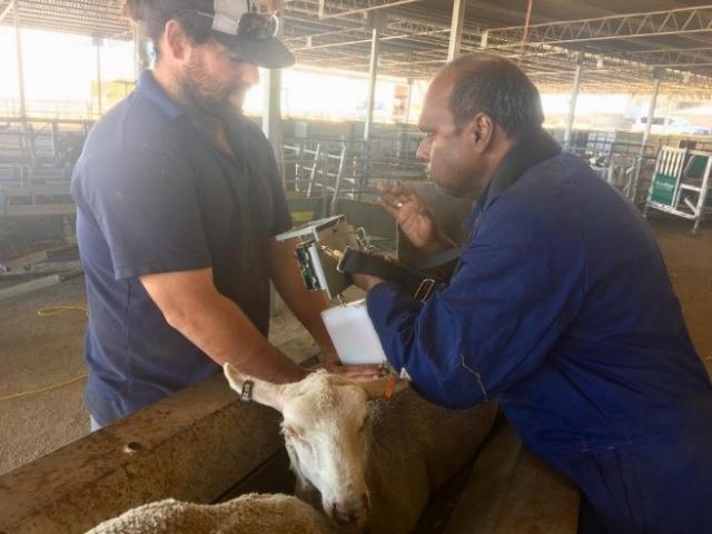
At this low power output the microwave measurement will not cause any damage to nor even be noticed by the animal. Lambs were immobilised in a race, the wool parted at the C-site and the VPA antenna was placed along the skin for scanning, to minimise the impact of wool on the microwave signal.
Microwave signals were then used to predict the ultrasound C-site fat depth and eye muscle depth using a range of machine-learning modelling methods including Support Vector Regression, Random Forest, and an Ensemble Stacking technique that combined both the Support Vector Regression, Random Forest and Partial Least Squares regression methods. All methods were trained using a 10-fold cross validation procedure, with R-square (R2) and root mean square error (RMSE) demonstrating the prediction precision (see Table 1).
R2 is a measure of the variation explained by the prediction model, with 1 being a perfect prediction, and RMSE is a measure of the error in the prediction model, with a smaller RMSE indicating the prediction based on the microwave C-site fat depth is close to the actual fat depth. The procedure was repeated with the inclusion of live weight in the predictive model (see Table 2).
Results and discussion
The lambs in the study demonstrated a wide range in live weight of 21 to 68kg; in ultrasound C-site fatness of 1 to 7.6mm and eye muscle depth of 9 to 34mm.
Microwave signals from the prototype hand-held device were able to predict ultrasound C-site fat depth measures with low to moderate precision and eye muscle depth with low precision. The Ensemble stacking method produced the best prediction of fat depth with R2 = 0.42 and RMSE = 0.736, and could provide a slightly more precise prediction of eye muscle depth (R2 = 0.27 RMSE = 3.685) than the Random forest method (Table 1, Figure 1).
Precision before accounting for live weight
|
| C-site Fat Depth | C-site Eye Muscle Depth | ||
|---|---|---|---|---|
| Modelling method | R2 | RMSE | R2 | RMSE |
| Partial least square regression (2 components) | 0.18 | 0.87 | 0.12 | 4.06 |
| Support vector regression | 0.36 | 0.78 | 0.22 | 3.81 |
| Random forest | 0.38 | 0.76 | 0.27 | 3.69 |
| Ensemble stacking | 0.42 | 0.74 | 0.27 | 3.68 |
RMSE = Root mean squared error; R2= R-square
Precision after accounting for live weight
| Modelling method (live weight included) | C-site Fat Depth | C-site Eye Muscle Depth | ||
|---|---|---|---|---|
| R2 | RMSE | R2 | RMSE | |
| Partial Least square regression (2 components) | 0.55 | 0.65 | 0.69 | 2.40 |
| Support vector regression | 0.58 | 0.63 | 0.70 | 2.38 |
| Random forest | 0.43 | 0.73 | 0.39 | 3.42 |
| Ensemble stacking | 0.61 | 0.60 | 0.70 | 2.35 |
RMSE = Root mean squared error; R2= R-square
The precision of microwave prediction of ultrasound C-site fat depth improved markedly when lamb live weight measures were included in the models (Table 2), with Ensemble stacking modelling of microwave data able to predict ultrasound fat depth with an R2 = 0.61 and RMSE = 0.604 (Table 2, Figure 2). Inclusion of lamb live weight in the modelling also substantially improved the ability of the Microwave scanner to predict ultrasound C-site eye muscle area, with an R2 = 0.70 and RMSE = 2.352 with the Ensemble stacking modelling.
Conclusion
These results demonstrate that the prototype handheld microwave scanner is capable of measuring C-site fat depth and eye muscle area with moderate precision, similar to the precision achieved for the prediction of C-site fat depth in carcasses reported in the January 2020 edition of the Ovine Observer. Of the different statistical methods tested the Ensemble stacking technique consistently produced the most precise predictions. This algorithm will be easily deployed within handheld microwave units to rapidly deliver fat depth predictions when measuring live animals in remote settings.
The microwave system predicted C-site fat depth with better precision than eye muscle depth, however the inclusion of live weight data improved eye muscle depth prediction to a greater extent than fat depth. This likely reflects the greater correlation between live weight and muscling or eye muscle depth compared to fat depth. Regardless, these greatly improved precision estimates demonstrate that the simultaneous use of a set of scales with a handheld microwave scanner can provide a quick and relatively precise estimate of C-site fat depth.
Small issues arose with microwave scanning in this experiment. This includes the technician’s fingers being too close to the microwave antennae when parting the wool for measurement, which is likely to have influenced the reflection of microwave signals. Additionally, the placement of the microwave antenna on the same location as the ultrasound scanning meant that some oil remained on the skin and may have influences the microwave measurements. Measures may be improved if the microwave antenna were placed parallel to the midline of the animals. These factors will be improved or investigated in further testing of the prototype scanner and may further improve the precision with which the system can predict fat and eye muscle depth.
The microwave system could be easily, safely and quickly used by untrained personnel to produce an objective measure of back fat in livestock that may help inform producers on when their stock are in optimal condition for slaughter.


