Livestock disease investigations protect our marketsAustralia’s ability to sell livestock and livestock products depends on evidence from our surveillance systems that we are free of particular livestock diseases. The WA Livestock Disease Outlook summarises recent significant disease investigations by Department of Primary Industries and Regional Development (DPIRD) vets and private vets that contribute to that surveillance evidence. |
Recent livestock disease cases in WA
Enterotoxaemia due to epsilon toxin of Clostridium perfringens type D
- Six of 150 five-month-old Merino lambs died in a 24-hour period. Five of the lambs were found dead, while one was observed showing signs of weakness and dull mentation. The affected lamb was presented to a private vet, but died shortly after arrival.
- Private vets and DPIRD vets performed a disease investigation and necropsy, which was subsidised through the Significant Disease Investigation Program.
- Enterotoxaemia due to epsilon toxin produced by Clostridium perfringens type D, commonly referred to as “pulpy kidney” was diagnosed as the cause of death. Histopathology showed perivascular leakage of high protein fluid around many of the vessels in the brain, and ileal contents were ELISA positive for enterotoxaemia epsilon toxin.
- Enterotoxaemia is typically seen in well grown lambs, particularly in the period after a feed change has occurred.
- Enterotoxaemia is best prevented by a vaccination program, though gradual change to higher quality feed can also reduce the risk of disease.
- Additional testing ruled out lead toxicity, which could cause similar signs of weakness and death in sheep (and cows), and is subject to regulatory activity if found in food producing animals.
Backyard poultry with facial swelling test negative for avian influenza
- A poultry owner contacted the Emergency Animal Disease hotline to report facial swelling in backyard poultry.
- Five of 60 chickens had swollen face wattles and combs.
- Thickened combs and wattles may be signs of the reportable diseases avian influenza and Newcastle disease.
- A DPIRD veterinarian conducted a disease investigation at the property, including collection of cloacal and choanal swabs from five affected chickens into viral transport medium and bacterial transport medium. The swabs in VTM were used to rule out AI and ND in the flock.
- The birds were vaccinated for the endemic poultry diseases Marek’s disease, infectious laryngotracheitis and infectious bursal disease.
- Avibacterium paragallinarum (formerly Haemophilus paragallinarum), the causative agent of the endemic poultry disease infectious coryza, was isolated from two of the five birds.
- Infectious coryza causes acute respiratory disease in poultry, usually restricted to upper respiratory tract infection, with nasal discharge and swollen head being the most common presenting signs.
- Most of the affected birds made a quick recovery.
Neurological cow tests negative for exotic disease
- Ante-mortem inspection at an abattoir detected an adult cow displaying neurological signs. The cow was circling and appeared blind. The animal was euthanised and the on-plant vet conducted a post-mortem.
- As the cow was displaying neurological signs consistent with the exotic disease bovine spongiform encephalopathy, the submitting vet supplied appropriate fixed and fresh brain and spinal cord samples for transmissible spongiform encephalopathy (TSE) exclusion testing. The test results were negative.
- The submitting vet observed purulent exudate on the brain, and a large abscess was seen in the midbrain when the pathologist was trimming the brain for histopathology.
- Histopathology confirmed the presence of abundant bacterial colonies in the abscess, and culture of the brain sample grew the opportunistic pathogen Trueperella pyogenes.
- This pathogen is one of the more common causes of sporadic brain abscesses in ruminants.
- To assure trading partners, markets and consumers that Australian animals and animal products are free of TSEs we need to conduct surveillance. The majority of this surveillance comes from post-mortem sampling of sheep and cattle that have neurological signs. Visit the TSE webpage for more information.
- Private veterinarians are eligible for rebates for submitting samples for TSE exclusion testing that meet the inclusion criteria. Laboratory submission forms are available on the forms page for vets.
Reminder: stay alert for E. canis

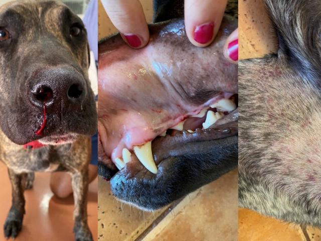
Dog with signs of E. canis. Epistaxis and oral and inguinal petechial haemorrhages can be seen. Bleeding is seen is some cases. [Photos courtesy of Dr John Beadle]
- Vets in the Kimberley, Pilbara, Gascoyne and Goldfields regions are reminded to remain vigilant for signs of Ehrlichia canis (ehrlichiosis) in dogs.
- Dogs become infected by E. canis when they are bitten by a brown dog tick (Rhipicephalus sanguineus) carrying the bacterium. Transmission does not occur directly between dogs.
- Clinical signs of E. canis in dogs are non-specific and commonly include lethargy, fever and inappetence. Other signs such as anaemia, haemorrhage, weight loss and ocular and nasal discharges have also been observed.
- DPIRD has been carrying out surveillance with the support of private vets in affected regions and working with other agencies to extend the messaging around E. canis across northern WA.
- It is recommended that owners living in or taking their animals to northern WA ensure their dogs are on an effective tick treatment.
- Dog owners should also check their dogs for ticks frequently and remove them if found.
- People moving their dogs out of the Kimberley must notify DPIRD and only move dogs if they are healthy and on a tick treatment. Details can be found on the Ehrlichiosis in dogs webpage.
In spring, watch for these livestock diseases
| Disease, typical history and signs | Key samples |
|---|---|
| Enterotoxaemia (pulpy kidney) in sheep
| Post- mortem
|
| Haemonchus (Barber’s pole worm) in sheep
| Ante-mortem
Post-mortem
|
| Polioencephalomalacia (PEM) in sheep and cattle
| Ante-mortem
Post-mortem
|
Recent cattle deaths a reminder to prevent livestock access to lead
- Several recent cattle deaths have occurred on two South-West properties as a result of lead ingestion. These cases are a reminder to consider lead toxicosis as a potential diagnosis in livestock displaying neurological signs.
- Animals affected by lead poisoning may become blind, unresponsive to their surroundings and bump into obstacles.
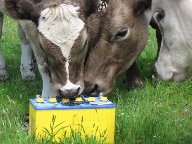
- DPIRD has reminded stock owners to remove or fence off items containing lead from grazing paddocks. Batteries, sump oil, paint, old machinery are all sources of lead and present a risk of both poisoning and residues.
- Preventing residues in meat and meat products is critical for human food safety and WA’s ongoing access to valuable export markets.
- For more information on preventing lead poisoning and residues in livestock visit the preventing residues in livestock webpage.

